CRISPR-Cas9 Genome Editing
A real window of the human genome on a ladder of brass and glass, with Cas9 clamped on it where its crystal structure puts it. Every score on the page is computed from published tables, on that sequence, as you watch.
Scroll
Twenty Letters in Three Billion
One set of human chromosomes holds about 3.1 billion base pairs. On the left is a metaphase chromosome, then the chromatin fibre unwinding out of it, and then what this model is really about: a length of double helix, and one address on it.
The address has two parts: twenty letters of target, and three letters of PAM right beside them. Twenty letters make a trillion combinations; asking for an NGG after them makes a chance hit in random sequence a matter of thousands to one.
This is a model built to the molecule’s own proportions: 3.4 ångström per base pair, a turn every 10.5 pairs, 20 ångström across.
3,088,286,401 bp (GRCh38: 24 chromosomes and the mitochondrion) · address 20 + 3 letters
The PAM First
Cas9 does not read the genome through. It collides with DNA and looks for an NGG: any base, then two Gs. If there is none it lets go and collides somewhere else.
Counting both strands, random sequence carries one NGG every 8 base pairs or so. Real sequence here is denser still. Every usable PAM in this window is lit on the ladder.
Arginines in the PI domain read those two Gs in the major groove. Only then does Cas9 prise the duplex open beside them.
1/16 per strand, about one every 8 bp counting both · the PAM is read in the major groove
Pairing, Base by Base
The duplex opens from the PAM end. The guide RNA’s twenty letters push in and pair with the target strand; the other strand is pushed out and loops around. The whole thing is an R-loop.
Pairing starts beside the PAM and runs away from it. Only when it reaches the far end does Cas9 take the shape that can cut.
On the ladder now: the target strand below, the guide RNA above it, and the displaced strand looping outside.
Pairing starts at the PAM · all 20 pairs before Cas9 cuts
Two Strands, Two Blades
Cas9 carries two nuclease domains. HNH cuts the strand the guide is paired with; RuvC cuts the strand that carries the PAM.
Both cuts fall at the third base pair upstream of the PAM, so the end they leave is blunt.
Having cut, Cas9 does not let go at once. The break waits there for the cell’s own repair.
HNH → target strand | RuvC → non-target strand · both 3 bp from the PAM · blunt
The Protein Taken Apart
This solid is the main chain of 1,368 amino acids, walked point by point through the coordinates of crystal structure 4UN3.
It falls into two lobes: the recognition lobe holds the RNA–DNA duplex, and the nuclease lobe carries both blades and the PAM-reading PI domain. A bridge helix joins them.
HNH does not face the strand it cuts. It swings across once all twenty pairs are made — one more thing keeping Cas9 from cutting where it should not.
REC | bridge helix | RuvC I–III | HNH | PI · domain limits from UniProt Q99ZW2
What It Will Forgive
One wrong letter does not always stop it. Far from the PAM, a mismatch is often ignored; within the dozen base pairs next to the PAM, it usually is not. That end is the seed.
The CFD score is a table of the activity measured for every mismatch at every position; several mismatches multiply. The score here is computed from that table, on these sequences.
Real off-targets have to be measured in cells. On the right are the sites GUIDE-seq found for this guide, with the reads as published; the scores beside them are the page’s own arithmetic.
CFD score (Doench et al. 2016) · seed: about the 10–12 bp next to the PAM
The Break Goes to the Cell
Cas9 only cuts. What follows is the cell’s: join the two ends, or copy from a template.
Joining is not random. Where the same short stretch occurs on both sides of the break, the sequence between them is often deleted whole; if what goes is not a multiple of three, the reading frame shifts and the gene is usually finished.
The other common outcome is one base gained: Cas9 sometimes leaves a single-base overhang, and filling it in copies the letter four positions upstream of the PAM.
The fourth tray is the template route: give the cell DNA carrying the change you want and it can rebuild the break from it. It is far less efficient.
Microhomology deletions and the out-of-frame share, computed here from Bae et al. 2014 · +1 copies the base at −4
Editing Without a Break
A double-strand break is a blunt instrument. Disable one blade or both, hang another enzyme on Cas9, and letters can be changed without severing anything.
A base editor carries a deaminase: on the short piece of single strand the R-loop exposes, it turns a C into a T, or an A into a G. No break, no template.
Prime editing puts a reverse transcriptase on a Cas9 nickase, and the guide RNA itself carries the sequence to be written in.
Base editing: C→T (Komor et al. 2016), A→G (Gaudelli et al. 2017) · prime editing (Anzalone et al. 2019)
Switching Fetal Haemoglobin Back On
Sickle cell disease and β-thalassaemia are both faults in one gene, HBB. The first approved CRISPR therapy does not repair it.
Before birth we run on a different haemoglobin, which a switch called BCL11A turns off afterwards. Casgevy takes a patient’s blood stem cells out and cuts BCL11A’s erythroid enhancer — inside the GATA1 binding site — so the switch fails and fetal haemoglobin comes back on.
Delivery is not one thing either: here the cells are taken out and Cas9 protein with its guide RNA is put in as a ribonucleoprotein. Viral vectors and lipid nanoparticles carry editors into the body instead.
The MHRA authorised it in November 2023; the FDA approved it for sickle cell disease in December 2023 and for transfusion-dependent β-thalassaemia in January 2024.
Casgevy (exa-cel) · the GATA1 site in BCL11A’s +58 erythroid enhancer · 2023
The Bench
Take a real gene, take a PAM, change a letter of the guide, and watch the score and the break change with it.
Try thisPut one wrong letter at position 2 of the guide, then one at position 18. Which costs more, and why is it the one next to the PAM?
What the model leaves out
The protein is the alpha-carbon main chain of 1,368 residues (PDB 4UN3), one solid per domain; the real thing is a mass of atoms and its surface is nothing like these smooth tubes. Where the crystal is disordered the model simply breaks. The DNA and RNA on the ladder are not the crystal’s coordinates: they are ideal B-form built to the measured figures (3.4 Å, 10.5 bp per turn, 20 Å), standing where the crystal’s duplex stood. In a real R-loop the RNA–DNA duplex is A-form, and the displaced strand does not loop as tidily as this. Bases are drawn as two lengths of rod, long for a purine and short for a pyrimidine; no atoms, no count of hydrogen bonds. The chromosome and the chromatin fibre in the first chapter stand for those scales rather than being the same molecule shrunk. A CFD score says how much cutting a mismatched site keeps, not that a cell will cut it; real off-targets are measured in cells. The microhomology arithmetic ranks deletion patterns; it does not give their frequencies.
Common misconceptions
CRISPR does not "cut a gene out and put a new one in": Cas9 only makes a break, and what the sequence becomes is the cell’s repair. Knocking a gene out works because the joining is sloppy, not because anything was replaced. The PAM is not on the guide RNA but on the DNA, and Cas9 reads it itself; without one there is no cut, which is part of how bacteria avoid cutting their own CRISPR record. Off-target cutting is not a matter of counting wrong letters: where they fall matters more than how many, and a mismatch next to the PAM costs far more. Casgevy does not repair the mutation that causes the disease; it edits a switch in another gene.
Sources
- Jinek, Chylinski, Fonfara, Hauer, Doudna & Charpentier (2012) Science: a dual-RNA-guided DNA endonuclease; HNH cuts the complementary strand, RuvC the non-complementary one; blunt ends 3 bp from the PAM
- Mojica et al. (2005) J Mol Evol: CRISPR spacers come from foreign genetic elements
- Barrangou et al. (2007) Science: CRISPR gives bacteria acquired resistance to viruses
- Cong et al. (2013) Science: multiplex genome engineering in human cells
- Mali et al. (2013) Science: RNA-guided human genome engineering
- Nishimasu et al. (2014) Cell: Cas9 with guide RNA and target DNA (PDB 4OO8)
- Anders, Niewoehner, Duerst & Jinek (2014) Nature: Cas9 bound to PAM-containing DNA (PDB 4UN3, the coordinates this model uses)
- RCSB PDB 4UN3
- UniProt Q99ZW2 (SpCas9): domain limits, active sites D10 and H840
- Sternberg et al. (2014) Nature: strand separation starts at the PAM
- Hsu et al. (2013) Nat Biotechnol: mismatch tolerance and the seed
- Doench et al. (2016) Nat Biotechnol: the CFD score and its mismatch and PAM tables
- Tsai et al. (2015) Nat Biotechnol: off-target sites and read counts from GUIDE-seq
- CRISPOR: Doench’s CFD calculator and its weight files
- Bae et al. (2014) Nat Methods: microhomology deletion patterns and the out-of-frame score
- Allen et al. (2019) Nat Biotechnol: what repair of Cas9 breaks produces, and how far it can be predicted
- Shen et al. (2018) Nature: inDelphi, predictable template-free editing
- Lemos et al. (2018) PNAS: the +1 insertion copies the base 4 nt from the PAM
- Taheri-Ghahfarokhi et al. (2018) Nucleic Acids Res: where single-base insertions fall and what they copy
- Komor et al. (2016) Nature: cytosine base editing
- Gaudelli et al. (2017) Nature: adenine base editing
- Anzalone et al. (2019) Nature: prime editing
- Canver et al. (2015) Nature: dissecting the BCL11A erythroid enhancer
- Wu et al. (2019) Nat Med: sgRNA-1617 cuts the GATA1 motif in the +58 enhancer
- Frangoul et al. (2021) NEJM: CTX001 in sickle cell disease and β-thalassaemia
- Frangoul et al. (2024) NEJM: exagamglogene autotemcel for severe sickle cell disease
- Locatelli et al. (2024) NEJM: exagamglogene autotemcel for transfusion-dependent β-thalassaemia
- MHRA (16 November 2023): the first authorised CRISPR therapy
- FDA (8 December 2023): approval for sickle cell disease
- EMA: Casgevy’s EU authorisation and product information
- Ensembl REST (GRCh38): the HBB and BCL11A sequence windows
- dbSNP rs334: HBB codon 6, GAG→GTG
- NCBI GCF_000001405.40 (GRCh38.p14): the assembly’s length
Data
The protein is the alpha-carbon trace of RCSB PDB entry 4UN3 (Anders, Niewoehner, Duerst & Jinek 2014), used under the PDB’s terms; the DNA and RNA on the model are idealised B-form, not the crystal’s coordinates. Sequence windows from Ensembl (GRCh38), cross-checked against the UCSC hg38 sequence API. The CFD mismatch and PAM tables are John Doench’s, as distributed with his CFD calculator and redistributed in CRISPOR (github.com/maximilianh/crisporWebsite); the microhomology arithmetic follows the source code published as Supplementary Figure 3 of Bae et al. 2014. Off-target sites and read counts from the supplementary tables of Tsai et al. 2015.
The text, pictures and models on this page are licensed CC BY-NC 4.0: free to use and adapt for teaching and other non-commercial purposes, with credit to world.lapla.si. Third-party material listed above keeps its own licence.
Follow the question
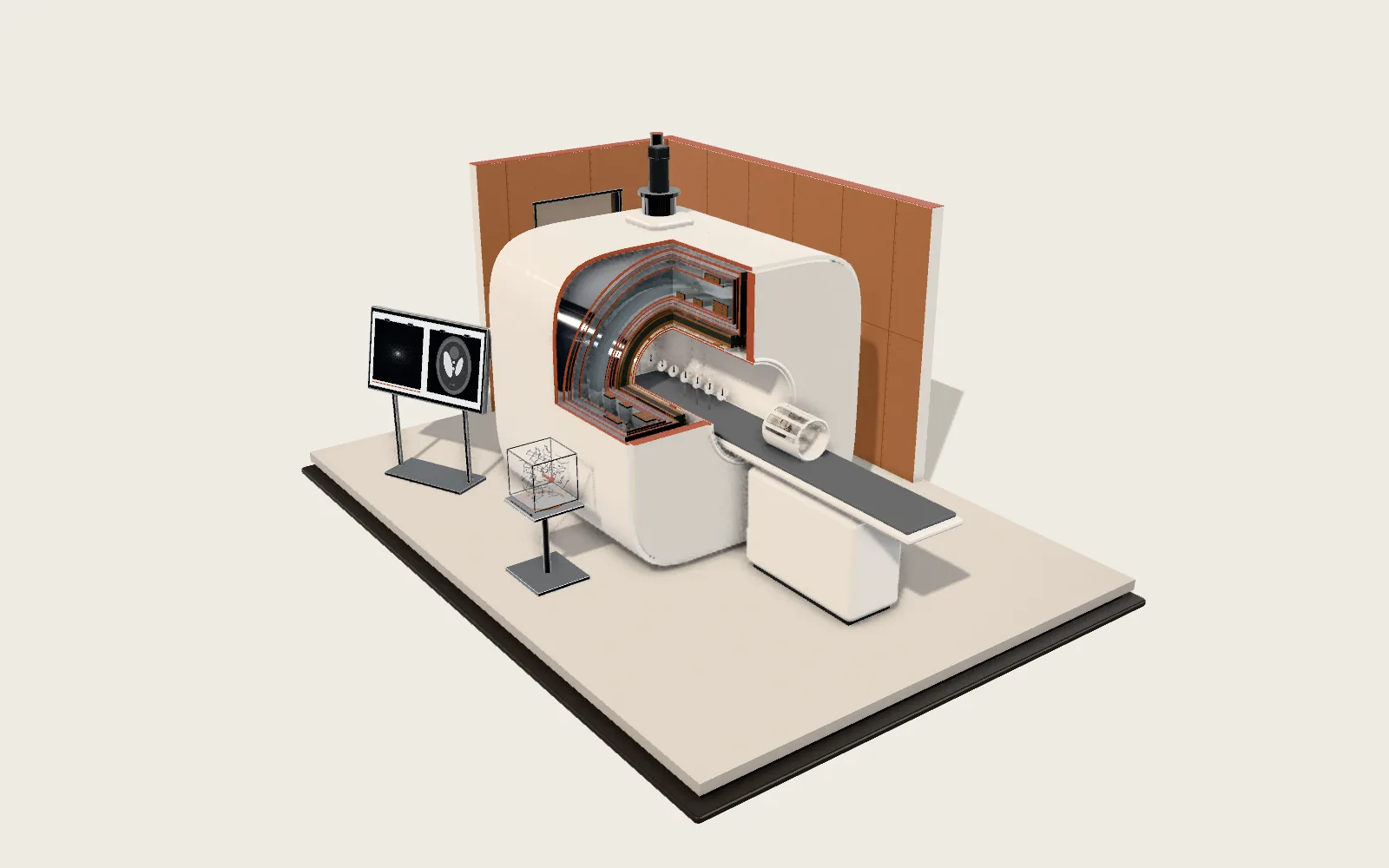 Plate F03MRI ScannerA clinical MRI scanner: a superconducting magnet, gradient coils and RF coils, one inside another. Hydrogen nuclei precess in the field; their signal fills k-space, and a Fourier transform turns it into an image.
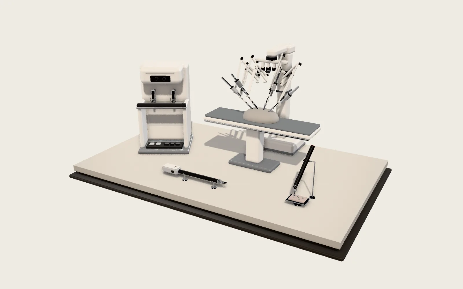 Plate F04Surgical RobotThe surgeon operates from a console nearby; the arms pivot each instrument about its incision. Hand motion is scaled down and stripped of tremor on its way to a tip 8 mm across.
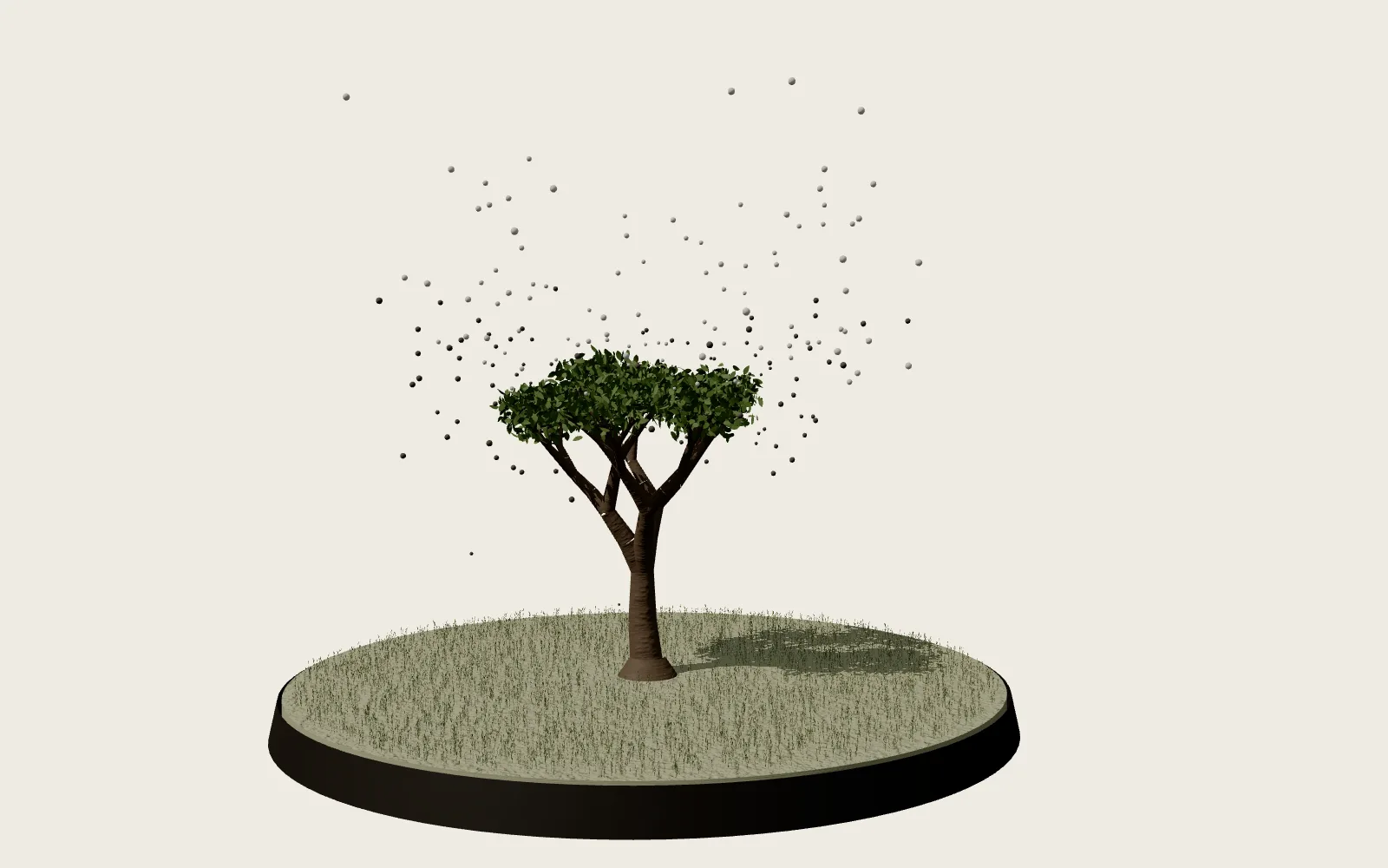 Plate 20The Breathing PlanetA tree’s wood is made mostly from air. Every summer the northern forests breathe in together, and the whole atmosphere holds less carbon dioxide.