How the Cochlea Takes Sound Apart
Deep in the inner ear is a tube coiled two and a half times, about 35 mm long when straightened. Sound sets a membrane inside it moving, and the motion runs inwards as a wave that swells to its highest at a place set by the frequency: high notes near the entrance, low notes at the far end.
Chapters · 11
- 01From the Ear Canal to the Inner Ear
- 02Getting Sound from Air into Fluid
- 03Uncoiling a 35-Millimetre Tube
- 04A Wave That Runs Inwards
- 05Every Frequency Has Its Place
- 06Hair Cells on the Membrane
- 07A Thread That Opens a Gate
- 08Hair Cells That Change Length
- 09A Trillionfold Range
- 10Cochlear Implants
- 11The Bench
Scroll
From the Ear Canal to the Inner Ear
The ear canal is about 2.5 cm long and 7 mm wide. It ends at the eardrum, about 9 mm across and 0.1 mm thick, shaped like a shallow funnel pointing inwards.
Behind the eardrum is the middle ear, a small air-filled space with a chain of three tiny bones: the handle of the malleus lies on the eardrum, the incus joins it, and the footplate of the stapes sits in the oval window of the inner ear. The stapes, about 3 mm tall, is the smallest bone in the body.
The inner ear is filled with fluid. The cochlea is the part that hears, about 9 mm across and coiled two and a half times; the three semicircular canals behind it sense balance.
Model: the ear about 100 times life size; the cochlea cut along its axis, its front half removed
Getting Sound from Air into Fluid
When sound in air meets water, about 99.9% of its energy bounces back; only about a thousandth gets in, a loss of about 30 decibels. Air is light and soft, water heavy and stiff: their impedances differ more than three thousandfold.
The middle ear wins most of this back. The eardrum’s working area is about 17 times the stapes footplate’s, so the same force on a smaller area makes 17 times the pressure; the malleus handle is about 1.3 times as long as the incus’ long process, a lever. Together that is ideally about 22 times the pressure, about 27 decibels.
Measured in human temporal bones, the middle ear’s gain is largest near 1 kHz, about 20 to 25 dB, and smaller at higher and lower frequencies.
The bones’ swing is exaggerated; the real motion is below a micrometre
Uncoiling a 35-Millimetre Tube
Straightened, the cochlea is a tube about 35 mm long, between 28 and 40 mm from person to person. It begins at the oval window and ends at the top of the coil.
Along its length the tube is divided in three. The upper channel is the scala vestibuli and the lower the scala tympani; they join at the far end, at the helicotrema, and hold perilymph. Between them runs the cochlear duct, the scala media, filled with endolymph rich in potassium and 80 to 100 millivolts positive.
The floor of the scala media is the basilar membrane, its roof the very thin Reissner’s membrane. When the stapes pushes the oval window in, the fluid cannot be compressed: it can only push the basilar membrane down and bulge out the thin membrane of the round window below.
The straightened tube is at the ear’s scale; its cross-section drawn 1.7 times larger and the basilar membrane 2.5 times wider. Reissner’s membrane is so thin that for the fluid the upper two channels act as one; the model leaves it out
A Wave That Runs Inwards
At the entrance the basilar membrane is narrow and stiff, about 0.1 mm wide; it widens and softens along the way to about 0.4 to 0.5 mm at the far end. The entrance is a hundred to ten thousand times stiffer than the far end.
Each push of the stapes starts a wave on the membrane that runs from the entrance towards the far end. As it goes it slows, shortens and grows, swells to its highest at one place, and dies away soon after.
Georg von Békésy first saw this travelling wave in cochleae from human cadavers, in the 1940s, and won the 1961 Nobel Prize in Physiology or Medicine. The peaks he saw were broad: in a living cochlea they are far sharper.
A 1 kHz tone. Heights on a decibel scale and enormously magnified: real motion is nanometres; time slowed about a thousandfold
Every Frequency Has Its Place
Where the wave peaks is set by its frequency. High notes peak near the entrance, low notes travel all the way to the far end. The basilar membrane is laid out like a keyboard, from 20,000 Hz down to 20 Hz.
In 1990 Donald Greenwood wrote the human map as a formula: f = 165.4 × (10^(2.1x) − 0.88), with x the position as a fraction of the length, measured from the far end. By it, 1 kHz peaks about 21 mm from the entrance.
Above 1 kHz each octave moves the peak 4.5 to 5 mm towards the entrance; low frequencies are packed closer. So we tell pitches apart roughly by ratio, not by difference.
The brass rule is engraved with each frequency’s place, from Greenwood’s formula
Hair Cells on the Membrane
On the basilar membrane sits the organ of Corti. On one side is a single row of about 3,500 inner hair cells; on the other, three rows of outer hair cells, about 11,000 to 12,000.
Each hair cell carries a bundle of stereocilia in three rows, each taller than the last, like a staircase. The tallest stereocilia of the outer hair cells are set into the tectorial membrane above.
When the basilar membrane bulges up, it and the tectorial membrane swing about different hinges, so the two slide a little against each other and push the bundles towards their tallest row. Of the roughly 30,000 auditory nerve fibres, about nine in ten connect to inner hair cells: what goes to the brain comes mostly from them.
A slice of the organ of Corti, about 3,500 times life size. The sliding is exaggerated
A Thread That Opens a Gate
Between neighbouring rows of stereocilia, from the tip of a shorter one to the side of the taller one, runs a thread about 150 nanometres long, the tip link, made of two cadherin proteins joined end to end.
When the bundle tips towards its tallest row the tip links tighten and pull open ion channels at the shorter stereocilia’s tips; when it tips back they slacken and the channels close. Potassium flows in from the endolymph, and the cell’s voltage rises and falls with it.
The gate is very fast, opening within tens of microseconds, and very sensitive: near the threshold of hearing the bundle moves only about a nanometre. Over less than about 100 nanometres the channels go from mostly closed to mostly open.
The tectorial membrane is lifted away so the bundles show; the tip links are drawn thicker; the brighter the colour, the larger the share of open channels
Hair Cells That Change Length
In 1985 William Brownell and colleagues found that outer hair cells shorten when their voltage rises and lengthen when it falls, by up to about 4% of their length. In 2000 Peter Dallos’ laboratory found the protein that does it and named it prestin. It works without chemical fuel, changing shape directly with the voltage, and keeps up with changes tens of thousands of times a second.
Moving with the sound, the outer hair cells pump energy into the basilar membrane just ahead of the peak. In a living cochlea this makes the peak 40 to 60 dB higher and much sharper; when the animal dies the gain is gone. Mice without prestin lose 40 to 60 dB of sensitivity.
The amplifier also leaks sound outwards. In 1978 David Kemp recorded echoes made by the cochlea itself in the ear canal, otoacoustic emissions. They are now widely used to screen newborn babies’ hearing.
The cells’ change of length is exaggerated. Readout: one faint tone along the membrane, with and without the amplifier
A Trillionfold Range
From the faintest sound we can hear to a painful one, the intensity spans about a trillionfold, 120 decibels. The cochlea fits this range by compressing it: for faint sounds the amplifier works at full gain, and the louder the sound, the less it adds. At the peak, every 10 dB more sound moves the membrane only 2 to 3 dB more.
Very loud sound damages hair cells: bundles are disarranged or broken and cells die. In people, as in other mammals, hair cells that die do not grow back. NIOSH recommends no more than 8 hours a day at 85 decibels, halving the time for every 3 dB more.
Some damage does not show on a hearing test. In a 2009 experiment on mice, noise raised the threshold for a while and it recovered within two weeks, yet in the worst-hit region about half the synapses between hair cells and nerve fibres were gone.
Outer-hair-cell bundles disarranged after noise (schematic); the tectorial membrane lifted away
Cochlear Implants
When hair cells are lost, the auditory nerve is often still there. A cochlear implant goes round the hair cells and stimulates the nerve directly: a slender electrode is slid through the round window into the scala tympani, one to more than one and a half turns.
Along the electrode are 12 to 22 contacts, depending on the maker. A processor outside the head splits sound into bands and sends high frequencies to contacts near the entrance and low ones to contacts further in: it borrows the cochlea’s own place map.
A few dozen contacts cannot replace thousands of hair cells, and the sound is coarse, but it is enough for many people to follow speech. By the end of 2019, about 737,000 cochlear implants had been registered worldwide.
The electrode is shown in the straightened tube, about 23 mm in; in a real cochlea it follows the scala tympani round the coil
The Bench
Frequency from 20 Hz to 20 kHz, level from 0 to 100 dB, a second tone if wanted, and the outer hair cells’ amplifier on or off. The peaks on the membrane and the curves in the readout come from the same model. Sound is off until started, and its loudness is only relative.
Try thisSet 1 kHz at 20 dB and switch the outer-hair-cell gain off: how many decibels does the peak drop, and where does it move? Compare again at 90 dB.
What the model leaves out
The travelling wave comes from a one-dimensional long-wave model: two fluid channels with a membrane between them whose stiffness falls along the tube to follow Greenwood’s map, driven by the stapes at the entrance and shorted by the helicotrema at the far end. The outer hair cells’ gain is added in the simplest way: just ahead of the peak the membrane’s damping is made negative, by an amount that shrinks as the level rises, giving about 40 dB more for faint tones and compression of about 0.28 dB per dB near the peak. It shows the travelling wave, the place map, the gain and the compression in outline, but it is not a fit to any one ear; the width and phase of the peak and the way two tones suppress each other are not modelled with care, and real peaks below about 1 kHz are less sharp. Heights are drawn on a decibel scale; real motion is nanometres. The three models are at different scales: the ear about 100 times life size, the straightened tube’s cross-section a further 1.7 times, the organ of Corti about 3,500 times.
Two common mistakes
The cochlea is not a row of separate tuning forks: the basilar membrane is continuous, the vibration travels along it as a wave from the entrance, and much of the sharpness of the peak comes from the living outer hair cells. And the outer hair cells mostly do not report sound to the brain; they amplify it. About nine in ten fibres carrying signals to the brain come from inner hair cells.
Sources
- Greenwood (1990): A cochlear frequency-position function for several species—29 years later
- Robles & Ruggero (2001): Mechanics of the mammalian cochlea, Physiol. Rev.
- Aibara et al. (2001): Human middle-ear sound transfer function and cochlear input impedance
- Puria (2003): Measurements of human middle ear forward and reverse acoustics
- Ulehlová et al. (1987): the length of the human basilar membrane
- Erixon et al. (2009): Variational anatomy of the human cochlea
- Liu et al. (2015): the human basilar membrane, width and structure
- Nobel Prize in Physiology or Medicine 1961: Georg von Békésy
- Ashmore (2008): Cochlear outer hair cell motility, Physiol. Rev.
- Spoendlin & Schrott (1989): the number of human auditory nerve fibres
- Wangemann (2006): endolymph and the endocochlear potential
- Fettiplace & Kim (2014): The physiology of mechanoelectrical transduction channels in hearing, Physiol. Rev.
- Kazmierczak et al. (2007): the tip link is made of two cadherins, Nature
- Corey & Hudspeth (1979): the speed of transduction
- Hudspeth (1989): How the ear’s works work, Nature
- Brownell et al. (1985): Evoked mechanical responses of isolated cochlear outer hair cells, Science
- Ashmore (1987): fast motile responses of isolated outer hair cells
- Zheng et al. (2000): Prestin is the motor protein of cochlear outer hair cells, Nature
- Frank, Hemmert & Gummer (1999): the frequency limit of outer hair cell motility
- Liberman et al. (2002): mice without prestin, Nature
- Kemp (1978): Stimulated acoustic emissions from within the human auditory system
- Penner & Zhang (1997): prevalence of spontaneous otoacoustic emissions
- Kujawa & Liberman (2009): synapse loss after noise, J. Neurosci.
- NIOSH: occupational noise exposure
- WHO and ITU: safe listening devices and systems (H.870)
- NIDCD: noise-induced hearing loss
- NIDCD: cochlear implants
- Dhanasingh & Jolly (2017): an overview of cochlear implant electrode arrays
Data
Place and frequency: Donald D. Greenwood, A cochlear frequency-position function for several species—29 years later (J. Acoust. Soc. Am. 87, 2592–2605, 1990). Cochlear length: B. Ulehlová, L. Voldřich and R. Janisch (Hear. Res. 28, 149–151, 1987); E. Erixon et al. (Otol. Neurotol. 30, 14–22, 2009). Basilar membrane width: W. Liu et al. (Cell Tissue Res. 360, 245–262, 2015). Middle-ear gain: T. Aibara, J. T. Welsh, S. Puria and R. L. Goode (Hear. Res. 152, 100–109, 2001); S. Puria (J. Acoust. Soc. Am. 113, 2773–2789, 2003). Cochlear mechanics: Georg von Békésy (Nobel Prize in Physiology or Medicine, 1961); Luis Robles and Mario A. Ruggero, Mechanics of the mammalian cochlea (Physiol. Rev. 81, 1305–1352, 2001). The travelling-wave model on the page is the one-dimensional long-wave model of the cochlea (after L. C. Peterson and B. P. Bogert, 1950), with the outer hair cells’ gain added as a local negative damping; tuned and drawn by this site. Hair cells: J. Ashmore, Cochlear outer hair cell motility (Physiol. Rev. 88, 173–210, 2008); H. Spoendlin and A. Schrott (Hear. Res. 43, 25–38, 1989); P. Wangemann (J. Physiol. 576, 11–21, 2006); R. Fettiplace and K. X. Kim, The physiology of mechanoelectrical transduction channels in hearing (Physiol. Rev. 94, 951–986, 2014); P. Kazmierczak et al. (Nature 449, 87–91, 2007); D. P. Corey and A. J. Hudspeth (Biophys. J. 26, 499–506, 1979); A. J. Hudspeth, How the ear’s works work (Nature 341, 397–404, 1989). The amplifier: W. E. Brownell, C. R. Bader, D. Bertrand and Y. de Ribaupierre (Science 227, 194–196, 1985); J. F. Ashmore (J. Physiol. 388, 323–347, 1987); J. Zheng et al., Prestin is the motor protein of cochlear outer hair cells (Nature 405, 149–155, 2000); G. Frank, W. Hemmert and A. W. Gummer (PNAS 96, 4420–4425, 1999); M. C. Liberman et al. (Nature 419, 300–304, 2002); D. T. Kemp (J. Acoust. Soc. Am. 64, 1386–1391, 1978); M. J. Penner and T. Zhang (Hear. Res. 103, 28–34, 1997). Noise and implants: S. G. Kujawa and M. C. Liberman (J. Neurosci. 29, 14077–14085, 2009); NIOSH, noise exposure limits; WHO–ITU H.870, safe listening devices; NIDCD, noise-induced hearing loss and cochlear implants; A. Dhanasingh and C. Jolly (Hear. Res. 356, 93–103, 2017). The models, the simulation and the words are the site’s own.
The text, pictures and models on this page are licensed CC BY-NC 4.0: free to use and adapt for teaching and other non-commercial purposes, with credit to world.lapla.si. Third-party material listed above keeps its own licence.
Follow the question
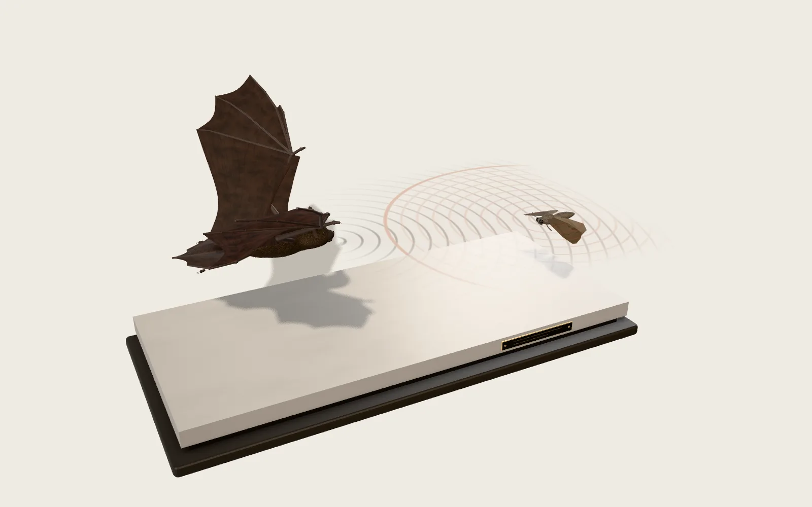 Plate 52Hearing a Moth in the DarkA bat hunts at night by its own voice. It sends out calls too high for us to hear and listens for the echo off a moth; how late the echo comes back tells it how far away the moth is.
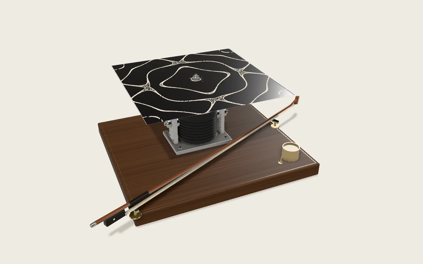 Plate 47Sand That Draws a SoundSprinkle sand on a metal plate and make it ring. The sand jumps away from the moving parts and gathers on the lines that stay still, drawing the shape of the vibration.