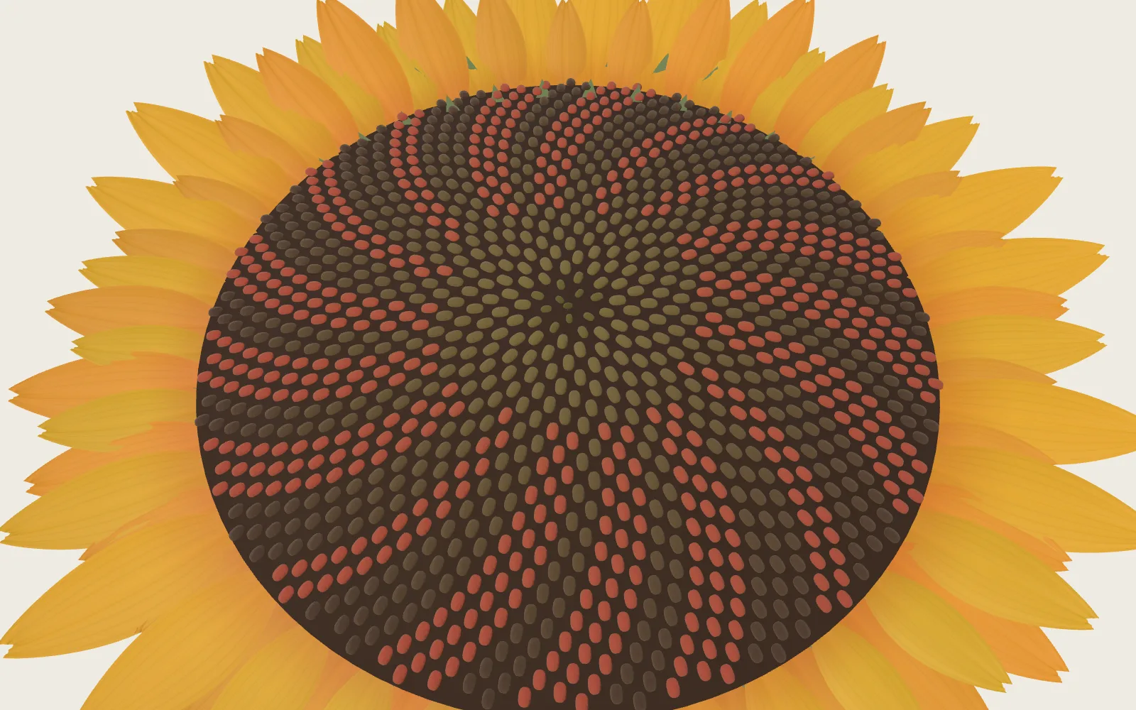
Mitosis in a Root Tip
Behind an onion root’s tip, cells keep dividing in two. A squashed, stained tip shows every stage at once; counting them tells how long each lasts.
Scroll
Four Zones at the Tip
A root’s tip wears a cap. It shields the dividing cells behind it; starch-filled amyloplasts settle to the lower side of its central columella cells and let the root sense gravity; its outermost cells keep coming away and new ones are added from inside.
Behind the cap is the meristem: small, nearly square cells with large central nuclei, where cells divide. Further back is the elongation zone, where cells lengthen fast and push the tip forwards; then the maturation zone, where the epidermis grows root hairs. It starts at the first root-hair bulges.
The zones don’t meet at neat lines: different files of cells stop dividing at different distances from the tip.
Root cap · meristem · elongation zone · maturation zone (root hairs)
Where Cells Divide
In the meristem the cells stand in files along the root. When a cell divides, a cross wall grows through its middle and the file gains a cell; the cells at the back are pushed into the elongation zone. In each cycle about half the meristem’s cells pass to the elongation zone and as many new ones are made.
At the meristem’s far tip is the quiescent centre, ringed by stem cells: they make new cap cells forwards and the files of cells backwards.
Dividing cells crowd the first short stretch of the root, so a school squash keeps only the last two or three millimetres.
Files of cells · the quiescent centre and stem cells · a squash takes the last 2–3 mm
Copy First, Then Divide
From one division to the next a cell goes through one cell cycle. Most of it is interphase: in G1 the cell grows, in S it copies its DNA, in G2 it gets ready to divide. Then comes mitosis, and last the cytoplasm splits in two.
In onion root tips at 21 °C one cycle was measured at about 23 hours, the S phase the longest at 12 hours and mitosis about 4 hours. Other studies found about 17–24 hours; temperature and method change the answer.
After S, each chromosome is made of two identical sister chromatids, held tightly together at the centromere.
Onion root tips at 21 °C: a cycle of about 23 hours · S about half of it · mitosis about 4 hours
The Chromosomes Condense
In prophase each chromosome coils up, thicker and shorter, until a light microscope can see it; the nucleolus disperses.
A plant cell makes one preparation an animal cell doesn’t: from G2 into prophase it builds a ring of microtubules and actin filaments against its membrane, the preprophase band. The band marks where the new wall will meet the old one; it comes apart as the nuclear envelope breaks down, but the place stays marked.
Onion chromosomes are large and few: 16 in a body cell (2n = 16), with about 16.4 billion base pairs in one set of eight, about five times a human set. That is why onion root tips are the usual material for watching division.
Chromosomes condense · the nucleolus disperses · the preprophase band (schematic)
Sixteen on One Plane
In prometaphase the nuclear envelope goes and the spindle’s microtubules reach in. Each chromatid has a kinetochore at its centromere; microtubules catch the kinetochores until the two sisters of each pair are tied to opposite poles.
In metaphase all 16 chromosomes lie on one plane midway between the poles, the metaphase plate. It is a place, not a structure.
Seed-plant cells such as the onion’s have no centrosomes: in prophase microtubules gather round the nuclear envelope, and when it breaks down they feed the spindle. The metaphase spindle is barrel-shaped, its poles not focused to a point. The cell waits until every kinetochore is attached before anaphase begins.
16 chromosomes · two chromatids each · the spindle (schematic)
The Copies Part
As anaphase begins, an enzyme cuts the cohesin that held each pair of sister chromatids together, and the two come apart, each now a chromosome of its own.
The microtubules tied to the kinetochores shorten and the chromosomes move towards the poles; the microtubules between the poles push them apart and the spindle lengthens. Each pole receives 16 chromosomes, the same as the cell had.
Cohesin cut · microtubules shorten · 16 to each pole
A Wall Grows Down the Middle
In telophase the two sets reach the poles and loosen, and a nuclear envelope forms round each: two nuclei.
A plant cell has a stiff wall and can’t pinch in from outside as an animal cell does; it builds a cell plate between the nuclei instead. The phragmoplast is an array of microtubules and actin filaments; vesicles from the Golgi carrying wall material travel along it to the midline and fuse into the plate. The phragmoplast turns from a cylinder into a ring and spreads outwards, and the plate grows from the centre out until it meets the old wall, where the preprophase band marked.
The plate becomes a new wall. One cell has become two, each with the same 16 chromosomes.
Nuclear envelopes return · the phragmoplast (schematic) · the cell plate grows outwards
Squashed and Stained
In a school practical the root tips are cut and fixed in a mixture of ethanol and acetic acid, which stops every cell where it is; warm dilute hydrochloric acid then loosens the middle lamella that cements the walls together; only the last two or three millimetres are kept, stained, covered with a coverslip and squashed to one layer of cells, and looked at at 400 times.
The usual stains are acetic orcein and toluidine blue; they darken the chromosomes, so dividing cells stand out at a glance.
A squash is a picture of the instant it was fixed: each cell is held at the stage it had reached. The order of the stages is pieced together from many cells, not watched in one.
Fix · soften in acid · stain · squash to one layer · look at ×400
Counting Out the Time
The share of cells caught dividing is the mitotic index. In control onion root tips it was measured at about 6–11 %.
When cells divide on their own, out of step with each other, the longer a stage lasts, the more cells a squash catches in it. So a stage’s length is about its share of the cells times the length of the cycle.
This page’s model takes the 23-hour cycle with 4 hours of mitosis at 21 °C and shares the 4 hours among the stages as measured at 15 °C: prophase about 99 minutes, metaphase (with prometaphase) about 33, anaphase about 25, telophase about 83.
It is only an approximation. On this model about 17 % of cells would be dividing; real counts find fewer, and with a 23-hour cycle that puts mitosis at only about 1.3–2.6 hours. Root tips also hold cells that have stopped dividing, and laboratories draw the line between stages differently: some count most cells in prophase, others in metaphase.
Control mitotic index about 6–11 % · the page’s model: prophase about 99 min, anaphase about 25 min
When the Split Goes Wrong
Now and then a pair of sister chromatids fails to separate and both go to the same pole: non-disjunction. One daughter cell gets an extra chromosome, the other is one short.
The spindle assembly checkpoint holds anaphase back until every kinetochore is attached, but some wrong attachments, such as one kinetochore tied to both poles, slip past it.
Colchicine binds the protein that microtubules are made of, so they cannot grow and the cell stops at metaphase; with no spindle, the chromosomes don’t line up but lie scattered through the cell. Since they never reach the poles, the cell’s chromosome number can double: in 1937 colchicine was first used to double the chromosomes of plants and make polyploids. Colchicine is highly poisonous.
Non-disjunction · the checkpoint · colchicine stops the spindle
The Cell Taken Apart
The dividing cell comes apart into five: the cells around it, its own wall and cytoplasm, its nuclear envelope breaking up, its 16 chromosomes and its spindle.
Taken apart, it is held in prometaphase: the envelope breaking up, the spindle’s microtubules reaching in. The cells around it fall back and it stands out alone. The part under the pointer lifts out alone and is named.
Cells around · wall and cytoplasm · nuclear envelope · chromosomes · spindle
Count a Field
This is the page’s model: the squash on the slide is drawn at random from the cycle above, each cell caught in a stage with a chance equal to that stage’s share of the cycle’s time, and every cell in the cycle. The page counts the cells in each stage in the field and works out the mitotic index and the length of each stage.
One field gives lengths that jump about; the more fields counted, the closer they come to the model’s own values (the fine lines on the chart). The lever sets how many fields are counted; each press of “New fields” samples and counts afresh.
The page’s model · a 23-hour cycle · about 140 cells a field
Try thisCount just 1 field and press “New fields” five times: between how many minutes does prophase jump? And with 20 fields?
What the Model Leaves Out
The root tip on the plinth is enlarged about eighty times and not to scale: there are far fewer, larger cells than in a real root, in files neater than real ones, and the cells on the lengthwise and cross cuts are a pattern drawn by position, not a section of any one root. The block behind is enlarged much further, with its front walls cut away; organelles are drawn only as a few grains. The chromosomes follow the onion’s 16, their lengths drawn; the spindle, the preprophase band and the phragmoplast are schematic lines, while real microtubules are far finer and far more numerous. The squash and the lab’s fields are drawn at random from a model: the stages’ lengths take the 23-hour cycle with 4 hours of mitosis at 21 °C (Matagne 1968) and share the mitosis among the stages as measured at 15 °C (Utrilla et al. 1993), with prometaphase counted as metaphase; it joins two sets of measurements, not one experiment.
Easy to Get Wrong
Most of the time a cell is not dividing but in interphase, which is why most cells in a squash are in interphase. A squash can’t show one cell dividing step by step: each cell is held at the instant it was fixed. Plant cells have no centrioles and don’t pinch in from outside; a cell plate grows from the middle. The metaphase plate is a place; the cell plate is the thing that grows. “The spindle fibres pull the chromosomes” is a shorthand: the microtubules tied to them shorten. Turning counts into times assumes the cells are out of step, all in the cycle, and classed the same way by everyone; counts from different laboratories differ a lot, so “prophase is always the most common” is not a rule. Some school exercises use a cycle of 80 minutes; that is a made-up number, and at room temperature an onion root tip’s cycle takes about 17–24 hours.
Common questions · 5
- What are the zones of a root tip?
Behind the cap is the meristem: small, nearly square cells with large central nuclei, where cells divide.
01Four Zones at the Tip ↑- What is the cell cycle?
From one division to the next a cell goes through one cell cycle.
03Copy First, Then Divide ↑- Do plant cells have centrioles?
Seed-plant cells such as the onion’s have no centrosomes: in prophase microtubules gather round the nuclear envelope, and when it breaks down they feed the spindle.
05Sixteen on One Plane ↑- How does a plant cell split in two?
A plant cell has a stiff wall and can’t pinch in from outside as an animal cell does; it builds a cell plate between the nuclei instead.
07A Wall Grows Down the Middle ↑- How do you work out how long each stage lasts from a squash?
When cells divide on their own, out of step with each other, the longer a stage lasts, the more cells a squash catches in it.
09Counting Out the Time ↑
Sources
- OpenStax: Biology 2e, 10.2 The Cell Cycle (Rice University, 2018)
- R. Matagne (1968): Duration of mitotic cycle and patterns of DNA replication in chromosomes of Allium cepa, Caryologia 21, 209 (a 23-hour cycle at 21 °C)
- L. Utrilla, M. I. Giménez-Abián and C. De la Torre (1993): Biology of the Cell 78, 235 (the stages’ timing at 15 °C)
- M. I. Giménez-Abián et al. (2004): Protoplasma 223, 191 (the onion root cycle; cold lengthens the phases evenly)
- M.-E. Ristea and O. Zarnescu (2024): Toxics 12, 194 (onion 2n = 16; the four zones; a control mitotic index)
- P. Ghosh, S. Roy and S. Ray (2025): Scientific Reports 15, 38837 (control counts by stage)
- T. Goh et al. (2022): Development 149, dev200593 (the root cap, columella, amyloplasts, border cells)
- M. A. Pacheco-Escobedo et al. (2016): Annals of Botany 118, 763 (the root’s zones, the quiescent centre)
- D. Francis, M. S. Davies and P. W. Barlow (2008): Annals of Botany 101, 747 (about half the meristem’s cells pass to the elongation zone each cycle)
- Royal Society of Biology, Practical Biology: Investigating mitosis in allium root tip squash
- SAPS: Root Tip Mitosis, technical and teaching notes (2019)
- H. H. Laughlin (1919): Carnegie Institution of Washington Publication 265 (a squash is a picture of the instant of killing)
- T. Potapova and G. J. Gorbsky (2017): The consequences of chromosome segregation errors in mitosis and meiosis, Biology 6, 12
- C. L. Asbury (2017): Anaphase A, Biology 6, 15
- M. Yamada and G. Goshima (2017): Mitotic spindle assembly in land plants, Biology 6, 6 (no centrosomes, the barrel-shaped spindle, the preprophase band)
- J. Choi, G. De Jaeger and H. S. Chung (2026): Cellular dynamics and molecular signaling networks of plant cytokinesis, Molecules and Cells 49, 100302 (the preprophase band, the phragmoplast, the cell plate)
- R. Finkers et al. (2021): Insights from the first genome assembly of onion, G3 11, jkab243 (2n = 16, about 16,400 Mb a set)
- NHGRI Talking Glossary: Base Pair (about 3 billion base pairs in one copy of the human genome)
- Y. Y. Leung, L. L. Yao Hui and V. B. Kraus (2015): Colchicine, update on mechanisms of action and therapeutic uses, Seminars in Arthritis and Rheumatism 45, 341
- W. F. Grant (1978): Environmental Health Perspectives 27, 37 (colchicine mitosis in onion root tips, as Levan described it in 1938)
- D. H. Touchell, I. E. Palmer and T. G. Ranney (2020): In vitro ploidy manipulation for crop improvement, Frontiers in Plant Science 11, 722 (colchicine first used to double chromosomes in 1937)
- P. Lara-Gonzalez, J. Pines and A. Desai (2021): Spindle assembly checkpoint activation and silencing at kinetochores, Seminars in Cell & Developmental Biology 117, 86
Data
The cell cycle of the onion root tip after R. Matagne (1968, 21 °C) and the timing of the stages of mitosis after L. Utrilla, M. I. Giménez-Abián and C. De la Torre (1993, 15 °C); the page’s model scales the one to the other (docs/plates/mitosis/computed-counts.py). Mitotic indices of onion root tips from M.-E. Ristea and O. Zarnescu (2024) and P. Ghosh, S. Roy and S. Ray (2025). The stages of the cell cycle after OpenStax Biology 2e and the reviews listed below; the school squash after the Royal Society of Biology’s Practical Biology and SAPS. Root zones after T. Goh et al. (2022) and M. A. Pacheco-Escobedo et al. (2016). The root, the block of cells, the squash and the lab are this page’s own model, not a photograph of any one root. Material presets and the model kit as on the site’s other model plates (public/looks/CREDITS.md).
The text, pictures and models on this page are licensed CC BY-NC 4.0: free to use and adapt for teaching and other non-commercial purposes, with credit to world.lapla.si. Third-party material listed above keeps its own licence.
Cite this plate
Laplace’s Mini World, Plate 61, “Mitosis in a Root Tip”, https://world.lapla.si/en/mitosis/, CC BY-NC 4.0, updated 2026-10-30.
Follow the question
 Plate F15CRISPR-Cas9 Genome EditingA ladder of DNA and a Cas9 built from its crystal coordinates: a guide RNA finds twenty letters among three billion, the PAM first, then base by base, and two domains cut one strand each.
 Plate 55Nerve ImpulseInside a nerve cell is about 70 millivolts below the outside. Pushed past threshold, voltage-gated sodium channels open and the membrane flips positive within about a millisecond, then potassium channels pull it back. The flip is made again, stretch by stretch, all along the axon; where the axon is wrapped in myelin it is made again only at the nodes of Ranvier, which is far faster.