AlphaFold: A Protein’s Shape from Its Sequence
A protein is a chain of amino acids, and the shape it folds into decides what it does. For decades a shape had to be measured by experiment, often over years. In 2020 AlphaFold, given only the sequence, predicted most protein shapes close to experimental accuracy. On the plinth is CI2, a small protein from barley seeds: the bronze ribbon is its prediction in the AlphaFold Database, the steel wire the structure measured by X-rays.
Scroll
A Chain of Amino Acids
A protein is amino acids joined one after another. There are 20 kinds, with different side chains: some avoid water, some carry a charge, some are tiny. The brass strip at the front carries all 84 letters of this one, one letter for each amino acid.
The chain folds up by itself: the water-avoiding side chains tuck inside, the charged ones stay outside. The shape decides the job. CI2 comes from barley seeds; it puts out a loop that fits into the active pocket of subtilisin and chymotrypsin and blocks both enzymes.
In 1973 Anfinsen summed it up: in the right surroundings, the sequence alone decides the shape. Could the shape then be worked out from the sequence? The question stayed open for nearly fifty years.
CI2 · UniProt P01053 · 84 amino acids · one letter per amino acid
The Folding Problem
A chain can take more shapes than anyone can count. In 1969 Levinthal pointed out that a protein cannot try them one by one. As the arithmetic is usually told: even with only 3 shapes per amino acid, 84 of them make about 10⁴⁰ combinations; trying ten trillion a second would take about 10²⁰ years. CI2 folds in water in about 20 milliseconds.
So shapes were measured. In 1958 Kendrew used X-ray crystallography to get the first three-dimensional structure of a protein. Today the Protein Data Bank holds about 260,000 experimental structures, while 150 million protein sequences are known.
Since 1994 structure prediction has had its own contest, CASP: every two years, the targets are structures just measured and not yet released, and the entrants get only the sequences.
3⁸⁴ ≈ 10⁴⁰ shapes · PDB 260,896 experimental structures · UniProtKB 150 million sequences (2026-10-18)
The Clue Evolution Left
The same kind of protein turns up in many species, with different sequences but much the same shape. Lining them up position by position gives a multiple sequence alignment. The nine rows above are nine members of CI2’s family, from different species.
When two amino acids touch in space and one changes, the other often changes with it, so the two still fit. Finding pairs of positions that change together across thousands of sequences hints at which ones touch.
These numbers are the site’s own, from the family’s 3,163 sequences in Pfam: of the 32 most strongly co-varying pairs, 22 really are in contact in the crystal structure. The thin brass rods join those 32 pairs. AlphaFold, too, starts by reading an alignment like this.
Pfam PF00280 · 3,163 sequences · 22 of the top 32 pairs in contact (beta carbons under 8 Å apart)
A Note for Every Pair
The body of AlphaFold 2 is called the Evoformer. It keeps a set of numbers for every pair of amino acids: 84 amino acids make an 84 × 84 grid, the pair representation. The alignment keeps its own.
Over 48 blocks the two trade information: the co-variation in the alignment is written into the pair grid, and the pair grid steers how the alignment is read. Attention lets each cell look at the cells that bear on it. A triangle update adds one more rule: if i touches j and j touches k, the distance from i to k is not free.
The plate at the back shows the distance of every pair, measured in this prediction; darker is closer. The dark patches away from the diagonal are parts far apart along the chain that sit together in space.
84 × 84 pairs · 48 Evoformer blocks · plate: alpha-carbon distance of every pair in the prediction, 0 to 20 Å
Building the Third Dimension
The structure module turns the pair grid into coordinates. It treats each amino acid as a small rigid body, all starting stacked at the origin and facing the same way; over 8 layers each one moves into place and turns to its angle, and then the side-chain angles are worked out.
The whole network runs four times, each pass taking the last one’s result as input. This is called recycling.
The chain on the plinth is doing something else, computed live on this page: starting as a flat string, it moves step by step by gradient descent, using only the predicted distance of every pair, until the distances match. The first AlphaFold, in 2018, built structures from predicted distances in this way; the second put this step inside the network.
Computed on this page: gradient descent from a flat chain to the predicted shape; the readout is how far this step is from the prediction
How Sure It Is
AlphaFold gives every amino acid a score from 0 to 100, called pLDDT: how accurate it expects the local structure there to be. The database paints four colours: dark blue above 90, light blue from 70 to 90, yellow from 50 to 70, orange below 50.
Nearly all of CI2’s body is dark blue; the first 20 are yellow and orange. In the crystal this stretch cannot be seen either: the first 18 amino acids of the mature protein have no fixed place. Very low pLDDT often means a stretch is loose to begin with.
The plate now shows the predicted aligned error: lined up on amino acid i, how many ångströms amino acid j is expected to be off. Darker green means the two positions are more reliable relative to each other.
pLDDT ≥ 90 · 70–90 · 50–70 · < 50 · the first 20 are below 70
Against the Experiment
The steel wire is CI2’s structure measured by X-rays in 1987, PDB entry 2CI2. With the prediction laid over it, the 65 positions they share are on average 0.80 Å apart, less than the length of a carbon–carbon bond (about 1.5 Å). That number is computed on this page.
This is no exam, though: 2CI2 was released long ago and was in AlphaFold’s training data. The real exam was CASP14 in 2020, with unreleased targets. AlphaFold’s median backbone error was 0.96 Å, against 2.8 Å for the next best method; on CASP’s GDT_TS score (100 is a perfect match) its median over the domains was 92.4.
From 2021 the predictions went into the public AlphaFold Database. By 2024 more than 214 million protein sequences had a predicted structure.
Prediction against crystal: alpha-carbon RMSD 0.80 Å (residues 20–84) · CASP14: 0.96 Å against 2.8 Å
What It Cannot Do
A prediction gives one still shape. Proteins move and change shape as they work; AlphaFold usually gives one of those shapes, and says nothing about how the chain folds.
Proteins seldom work alone. AlphaFold 2 works on protein chains. AlphaFold 3, in 2024, takes proteins, DNA and RNA, small molecules, ions and modified residues together, and makes the atom coordinates directly with a diffusion module.
Its paper names the limits too: in loose regions it sometimes draws order that is not there, and its structures do not always respect chirality. A prediction is a hypothesis; before it is used for experiments or drugs, it often needs checking.
AlphaFold 3 (2024): proteins · nucleic acids · small molecules · ions · modified residues
The Nobel Prize
The 2024 Nobel Prize in Chemistry went half to Demis Hassabis and John Jumper, for protein structure prediction, and half to David Baker, for computational protein design: the other way round, choosing a shape first and working out a sequence that folds into it.
From Anfinsen’s 1973 statement that sequence decides shape to computing the shape from the sequence as an everyday tool took less than fifty years.
Nobel Prize in Chemistry 2024
The Bench
The slider is the number of gradient-descent steps: the chain starts as a flat string, or as a random coil, and moves step by step towards the predicted distances. The readouts give how far this step is from the prediction and from the crystal structure. The pLDDT colours, the crystal structure and the co-varying pairs can be switched on.
Try thisTurn on the pLDDT colours, then set the crystal structure inside. Why does the steel wire not follow the orange and yellow stretch?
What the model leaves out
The ribbon is CI2’s prediction in the AlphaFold Database (AF-P01053-F1, version 6), drawn from each amino acid’s alpha carbon only; the extent of the helix and the strands is taken from the crystal structure 2CI2. A ribbon is a drawing convention; atoms do not look like this. The chain folding from flat is not AlphaFold’s computation but this page’s distance geometry: a fit to all the predicted distances, untangled first in four dimensions and then pressed back into three. It shows only that knowing every pair’s distance means knowing the shape. The co-variation is the site’s own, from Pfam’s alignment with a pseudo-likelihood model, not the alignment AlphaFold uses (it searches UniRef90, BFD, MGnify and others).
Common misunderstandings
Saying AlphaFold “solved protein folding” is loose: it predicts the folded shape, not the folding process, nor every state a protein takes in a cell. A high pLDDT means the local structure is reliable, not that the placing of two domains relative to each other is; that is what the predicted aligned error is for. AlphaFold does not compute shapes from the laws of physics either: it learned the link between sequence and shape from decades of experimental structures.
Common questions · 4
- What makes protein folding hard?
A chain can take more shapes than anyone can count. In 1969 Levinthal pointed out that a protein cannot try them one by one.
02The Folding Problem ↑- How can evolution reveal which amino acids touch?
When two amino acids touch in space and one changes, the other often changes with it, so the two still fit.
03The Clue Evolution Left ↑- What do the four pLDDT colours mean?
The database paints four colours: dark blue above 90, light blue from 70 to 90, yellow from 50 to 70, orange below 50.
06How Sure It Is ↑- How accurate was AlphaFold at CASP14?
AlphaFold’s median backbone error was 0.96 Å, against 2.8 Å for the next best method
07Against the Experiment ↑
Sources
- Jumper et al. (2021): Highly accurate protein structure prediction with AlphaFold, Nature 596:583–589 (and supplementary information)
- Jumper et al. (2021): Applying and improving AlphaFold at CASP14, Proteins 89:1711–1721 (median domain GDT_TS 92.4)
- Kryshtafovych et al. (2021): Critical assessment of methods of protein structure prediction (CASP)—Round XIV, Proteins 89:1607–1617
- Abramson et al. (2024): Accurate structure prediction of biomolecular interactions with AlphaFold 3, Nature 630:493–500
- Senior et al. (2020): Improved protein structure prediction using potentials from deep learning, Nature 577:706–710
- Varadi et al. (2022): AlphaFold Protein Structure Database, Nucleic Acids Res. 50:D439 (the four pLDDT bands)
- Varadi et al. (2024): AlphaFold Protein Structure Database in 2024, Nucleic Acids Res. 52:D368 (over 214 million)
- AlphaFold DB: AF-P01053-F1 (CC BY 4.0)
- McPhalen & James (1987): crystal structure of CI-2, Biochemistry 26:261–269; PDB 2CI2
- Jackson & Fersht (1991): Folding of chymotrypsin inhibitor 2. 2, Biochemistry 30:10436 (folding rate 53 s⁻¹)
- UniProt P01053: Subtilisin-chymotrypsin inhibitor-2A, barley
- Anfinsen (1973): Principles that govern the folding of protein chains, Science 181:223
- Levinthal (1969): How to fold graciously
- Zwanzig, Szabo & Bagchi (1992): Levinthal’s paradox, PNAS 89:20
- Kendrew et al. (1958): A three-dimensional model of the myoglobin molecule, Nature 181:662
- CASP (Prediction Center)
- Pfam PF00280 (InterPro; Blum et al. 2025, Nucleic Acids Res. 53:D444)
- Marks et al. (2011): Protein 3D structure computed from evolutionary sequence variation, PLoS ONE 6:e28766
- Morcos et al. (2011): Direct-coupling analysis of residue coevolution, PNAS 108:E1293
- Ekeberg et al. (2013): Improved contact prediction in proteins: using pseudolikelihoods to infer Potts models, Phys. Rev. E 87:012707
- Nobel Prize in Chemistry 2024, press release
- RCSB PDB statistics; UniProtKB release 2026_03 statistics
Data
The predicted structure, its pLDDT and its predicted aligned error are AlphaFold DB entry AF-P01053-F1, version 6 (Jumper et al. 2021; Varadi et al. 2024), used under CC BY 4.0. The experimental structure is RCSB PDB entry 2CI2 (McPhalen & James 1987), CC0 1.0. The family alignment is Pfam PF00280 (Potato inhibitor I family) from InterPro (Blum et al. 2025), CC0 1.0. The co-evolution scores, the superposition and the fold on the bench are the site’s own, computed by docs/plates/alphafold/coevo.py and src/topics/alphafold/science.js.
The text, pictures and models on this page are licensed CC BY-NC 4.0: free to use and adapt for teaching and other non-commercial purposes, with credit to world.lapla.si. Third-party material listed above keeps its own licence.
Cite this plate
Laplace’s Mini World, Plate F19, “AlphaFold: A Protein’s Shape from Its Sequence”, https://world.lapla.si/en/alphafold/, CC BY-NC 4.0, updated 2026-10-18.
Follow the question
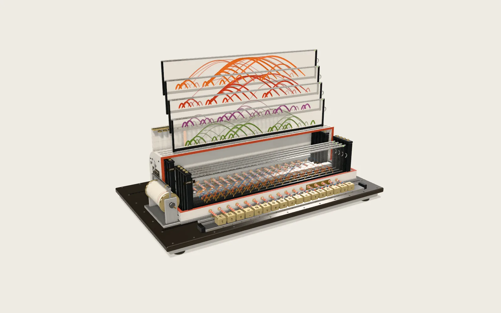 Plate F13Attention in TransformersA two-layer, eight-head Transformer reads a sentence in your browser, one character at a time, and guesses the next. Each thread is one act of attention; its brightness is the weight.
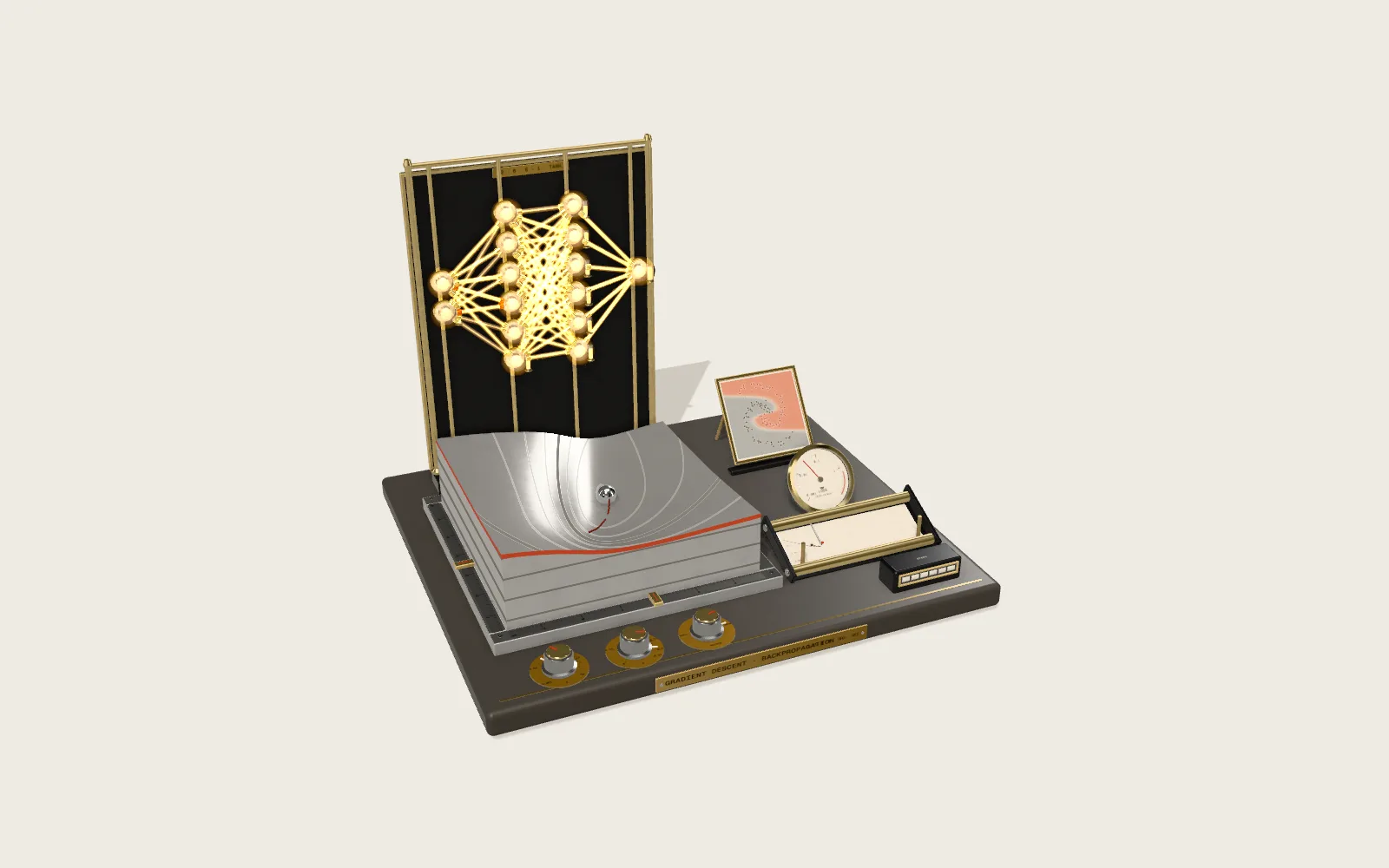 Plate F18How a Neural Network LearnsAn aluminium relief milled to a network’s real error, a steel ball that steps down it by the real gradient, and a small network in glass and brass: light runs forward with the numbers and back with each weight’s share of the blame.
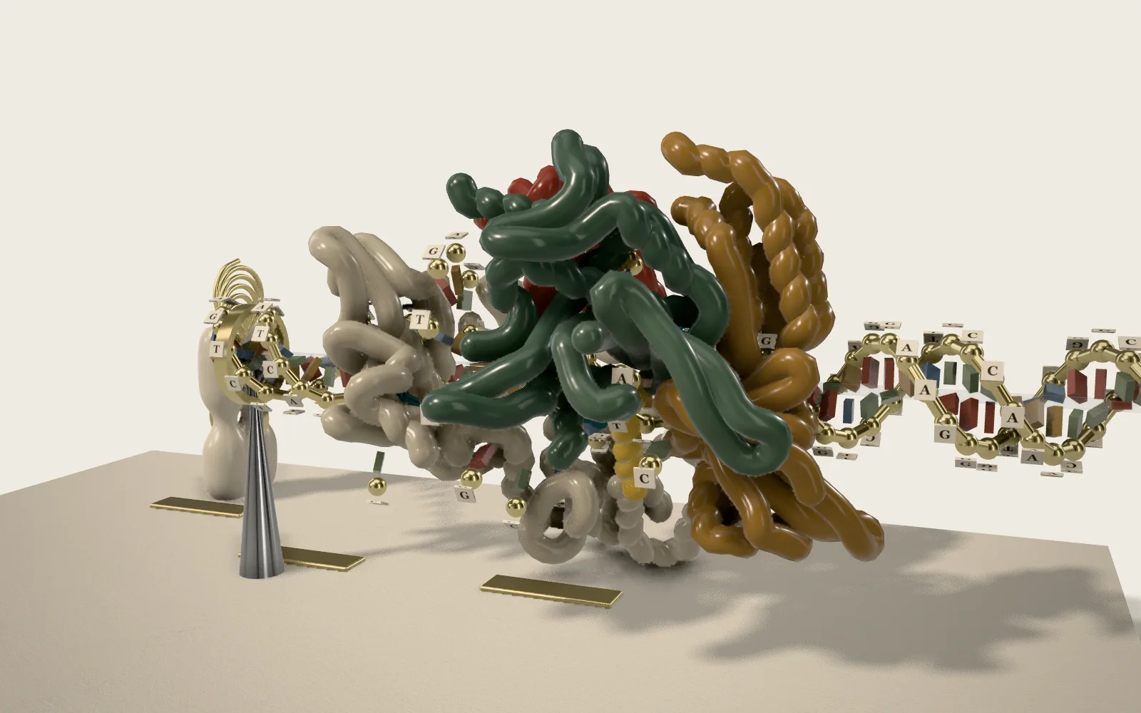 Plate F15CRISPR-Cas9 Genome EditingA ladder of DNA and a Cas9 built from its crystal coordinates: a guide RNA finds twenty letters among three billion, the PAM first, then base by base, and two domains cut one strand each.
 Plate 44Pictures Out of NoiseMany AI image generators start from random static and remove it, one step at a time. A small network does the same in your browser, and a cloud of dots slowly condenses into a knot.