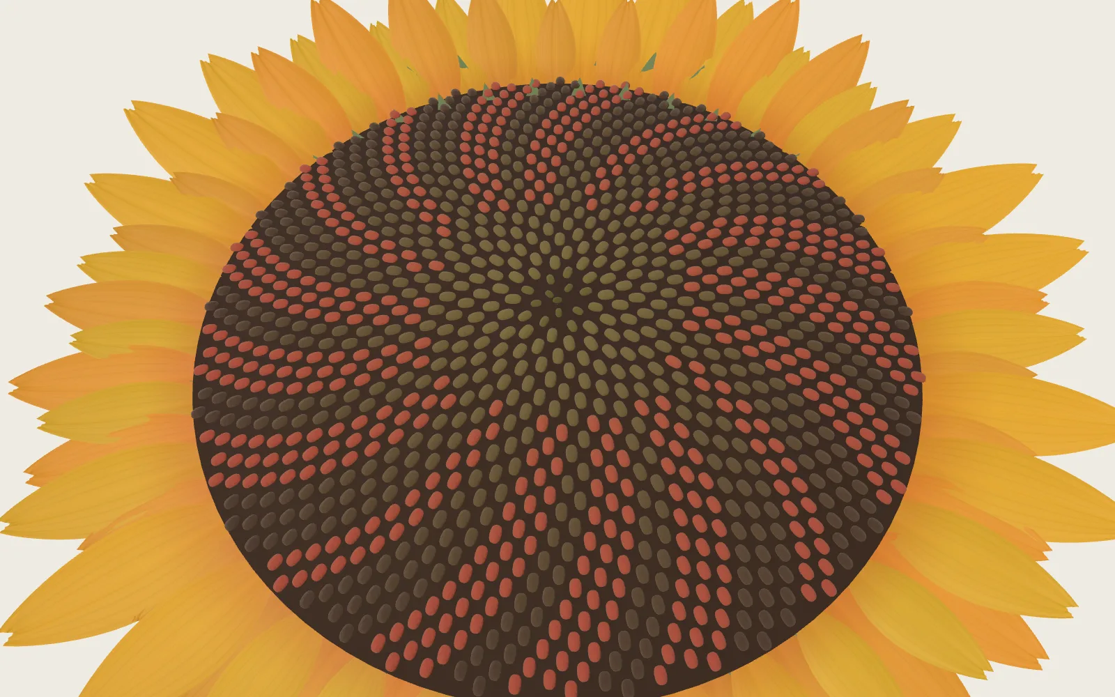
Across the Cell Membrane
A cell membrane is two layers of lipid, under 5 nm thick. Water seeps through; salt and sugar need a protein. In strong salt, a red onion cell’s purple pulls away from its wall.
Chapters · 12
Scroll
Two Layers, Under Five Nanometres
The body of a cell membrane is two layers of lipid molecules. Each has a head that likes water and two oily tails; in water the tails hide inwards and the heads face out, and the molecules settle into two layers by themselves.
A pure phospholipid bilayer is about 4.5 nm thick with its heads, and its oily core about 2.7 nm. Membranes from rat liver cells measure 3.6–4.3 nm from the phosphates on one side to those on the other.
A square micrometre of membrane holds a few million lipids. They jostle constantly within their own layer, but crossing to the other is slow: without an enzyme, half of them take 6.5–15 hours to flip.
On the plinth is a patch 16 nm wide, enlarged about 32 million times (computed).
Pure phospholipid: core about 2.7 nm, about 4.5 nm with heads · cell membranes: phosphate to phosphate 3.6–4.3 nm
What Gets Through on Its Own
The barrier is the oily core. Small uncharged molecules such as oxygen and carbon dioxide barely notice it; water is polar but seeps through slowly; urea and glycerol are a few hundred times slower again.
Ions hardly get through at all: across an artificial bilayer, sodium and potassium have permeabilities of about 5 × 10⁻¹⁴ cm/s, some ten orders of magnitude below water. Carrying a charge into oil costs too much. Glucose is large and polar, and does not get through either.
For ions and sugars to move in and out, a cell needs proteins in its membrane.
Artificial bilayers, cm/s: O₂ 23–125 · CO₂ 0.35 · water about 0.003 · urea about 0.000004 · Na⁺ about 5 × 10⁻¹⁴
Water in Single File
The water channel aquaporin-1 is abundant in red blood cells and kidney tubules. It is four identical subunits, each with a narrow pore of its own; the model is cut open along one of them.
At its narrowest the pore is about 2.8 Å, barely wider than a water molecule; along some 20 Å in the middle, waters can only pass one behind another. Two NPA motifs sit at the middle, where the field turns each water round; the electrostatic barrier there is what mainly keeps protons out, so the pore passes water but not H⁺.
From its measured unit permeability, each subunit can pass up to about 3 billion water molecules a second. How many actually flow depends on the osmotic difference: with the two sides equal, the water shuffles back and forth with no net flow.
AQP1 (PDB 7UZE) · narrowest about 2.8 Å · waters in single file (schematic)
An Egg That Burst
In 1988 researchers purified a 28 kDa protein from red cell membranes: 120,000–160,000 copies on each cell, and no one knew what it did.
In 1992 they injected its RNA into eggs of the African clawed frog, waited 3 days for the protein to appear in the egg membrane, and moved the eggs into a hypotonic solution. Within 30 seconds the eggs began to swell; after 3 minutes they burst. Eggs injected with water alone barely changed. A mercury salt blocked the effect, reversibly.
It was the first water channel to be recognised, later named AQP1, and Peter Agre shared the 2003 Nobel Prize in Chemistry for it. From above, the four subunits make a square, each with its own pore; the central axis is not a water path.
120,000–160,000 per red cell · eggs swell in 30 s and burst in 3 min · four subunits, four pores
A Pocket That Turns
Glucose enters red cells through GLUT1. GLUT1 is not an open pore: a sugar enters a pocket in its middle from one side, the protein changes shape, the pocket opens to the other side, and the sugar leaves.
It moves sugar only down the concentration difference and uses no energy: facilitated diffusion. One GLUT1 carries about 1,200 glucose molecules a second; an open potassium channel passes hundreds of millions of ions a second.
The model’s GLUT1 comes from an inward-open crystal (PDB 6THA); the sugar ring in its pocket is the glucoside of a detergent used to grow the crystal, and the outward-open shape is known from its close relative GLUT3. Moving things against the difference takes energy, which is a pump’s job: the sodium–potassium pump moves 3 sodium ions out and 2 potassium ions in for each ATP (see “Nerve Impulse”).
GLUT1 (PDB 6THA, inward-open) · about 1,200 glucose a second · the sugar’s path is a schematic
Which Way the Water Goes
When one side of a membrane holds more dissolved particles that cannot cross it, water flows, on balance, towards that side: osmosis. What counts is the number of particles, not what they are: sodium chloride splits into a sodium ion and a chloride ion, so each counts twice.
Holding the flow back takes extra pressure on the side with more particles: the osmotic pressure. In a dilute solution π = cRT. With the measured osmotic coefficients of sodium chloride, 0.9 % salt at 25 °C gives about 0.71 MPa and 3 % about 2.3 MPa, some 23 atmospheres (computed).
In 1877 Pfeffer first measured osmotic pressure, with a membrane that let water through but not sugar. Van ’t Hoff found that a dilute solution’s osmotic pressure equals the pressure of the same number of molecules as a gas, and in 1901 won the first Nobel Prize in Chemistry.
More solute above (schematic) · 0.9 % NaCl about 0.71 MPa · 3 % about 2.3 MPa (25 °C, computed)
Why Drips Are 0.9 %
Blood plasma runs at about 275–295 mOsm/kg. Saline of 0.9 % holds 9 g of sodium chloride per litre: 308 mOsm/L if each splits into two particles, about 286 mOsm/kg as measured, close to plasma. That is why it is called isotonic and used in drips. It holds only sodium and chloride, so it is not the same as plasma.
Red blood cells have no wall. In weaker salt, water flows in and they swell into spheres: at about 0.45–0.53 % the most fragile begin to leak haemoglobin, and by about 0.27–0.33 % nearly all have (the figures shift with temperature). The haemoglobin mostly leaks out through a stretched membrane; the membrane need not break apart.
In strong salt, red cells lose water and shrink into spiky little balls.
0.9 % NaCl: 308 mOsm/L nominal, about 286 mOsm/kg measured · plasma 275–295 mOsm/kg
A Cell with a Wall
A plant cell has a wall outside its membrane: a cross-linked mesh of pectin, cellulose and other polymers, stiff and full of pores that let water and salt through. What chooses what goes in and out is the membrane inside it.
The outer skin of a red onion scale is a single layer of long cells; onion epidermal cells that have been measured are about 0.4 mm long and 50–120 µm wide. The purple is anthocyanin, dissolved in the sap of the vacuole. In a mature plant cell the vacuole often fills over nine tenths of the cell, and the cytoplasm is a thin layer pressed to the membrane.
In fresh water the cell takes up water and presses its membrane against the wall: turgor, about 0.3–0.7 MPa in onion skin, depending on how it is measured. The wall holds against it, which is why a plant cell in fresh water does not burst. The cells on the plinth are enlarged about 160 times (computed).
Red onion epidermis · turgor about 0.3–0.7 MPa · vacuole often over nine tenths of the cell
The Purple Pulls Back
Drop strong salt water at one edge of the cover slip and draw the water off at the other with blotting paper, and the salt water flows past the cells. With more particles outside than in the sap, water leaves through the membrane: the vacuole shrinks, the purple looks deeper, and the protoplast pulls away from the wall. This is plasmolysis.
The wall hardly moves, and what fills the space between wall and protoplast is the salt water from outside, not air. Where the membrane leaves the wall, fine threads often still tie it on: Hechtian strands.
In strong salt the protoplast can be seen leaving the wall within tens of seconds, and settles in minutes to half an hour, faster in stronger solutions. In 1877 de Vries named the effect plasmolysis, and later used it to find the strength of solution that matches the sap.
3 % NaCl: the protoplast shrinks to about 41 % (this page’s model) · salt water between wall and protoplast
Back in Fresh Water
Back in fresh water, with fewer particles outside, water flows back into the cell and the protoplast swells against the wall again. Tobacco cells finish plasmolysing in 3–5 minutes and recover their shape 10–20 minutes after going back into their medium.
Only a living cell can go back and forth like this. Living cells survive more than a day plasmolysed; a cell whose membrane has failed cannot hold its sap, so it no longer shrinks and swells. Dried too fast and then flooded, the membrane may not have enough area and the protoplast bursts, which is why researchers bring cells back through a weaker solution first.
Salting cabbage works the same way: water flows out of the leaves into the brine, salt slowly moves in, and the leaves lose water, soften and shrink.
Tobacco cells: plasmolysed in 3–5 min, recovered in 10–20 min · only living cells recover
A Patch of Membrane, Taken Apart
The model comes apart in five: the outer leaflet, the inner leaflet, aquaporin-1, the glucose carrier GLUT1, and the file of water in the pore. The outer leaflet lifts off, and the two proteins and the water file come forward.
Both proteins’ surfaces are computed from Protein Data Bank atoms and set in the membrane at the depth the OPM database gives. Rest the pointer on a part and it is lifted out on its own and named.
Outer leaflet · AQP1 · GLUT1 · water file · inner leaflet
Laboratory: Salt Outside
This is the page’s model: set the strength of the salt water outside the onion cells. Weaker than the sap, the cells stay full; past incipient plasmolysis the protoplast shrinks until the osmotic pressures inside and out match; back in fresh water it swells again.
The sap is set at 0.8 MPa (onion skin measures about 0.5–1 MPa), the part with no water to give at 10 %, and 95 % of a change takes about 3 minutes, with a minute passing in about 6 seconds on the page. Each move of the lever carries on from the cells’ present size; “Start again” starts them full.
This page’s model · plasmolysis from about 1.0 % · about 41 % at 3 %
Try thisTake the salt outside from 0 to 3 %: at about what strength does the purple part start to pull away from the wall? Back in fresh water, does it swell to its old size?
What the model leaves out
The patch on the plinth is 16 nm wide, enlarged about 32 million times (computed). Both proteins’ surfaces are computed from their atoms: AQP1 from PDB 7UZE (human red cell membrane, cryo-EM, 2.4 Å), GLUT1 from PDB 6THA (2.4 Å). Each surface treats every atom as a soft ball, an approximation; hydrogens are not drawn, and the pore is found from the atoms the way the HOLE program does it. The lipids are drawn: one head and two tails, at the real thickness, all of one kind, with no cholesterol or sugar chains; real membranes are more crowded with protein. The waters in the pore, the small molecules, ions, glucose and solute particles are schematics, not simulations of where they are or how fast they go. The onion cells, the bottle and the slide are at their own scales. The lab is the page’s model: the sap’s osmotic pressure, the part with no water to give and the time to settle are chosen parameters, and salt slowly entering the cells or membranes giving way are left out.
Easy to get wrong
Water does not cross only through water channels: without AQP1 it still seeps through the lipid, only much more slowly; in human red cells roughly nine tenths of the osmotic water flow goes through AQP1, and people without AQP1 have no symptoms in daily life. Osmosis is not salt pulling water: any particle that cannot cross does the same, and what counts is their number. “3 billion a second” is the limit per subunit, not per tetramer, and the actual flow depends on the osmotic difference. GLUT1 uses no energy and moves sugar only down the difference; moving against it takes a pump. A plasmolysed cell is not a dead one: a living cell recovers in fresh water, a dead one does not. The space between wall and protoplast holds the outside salt water, not air. Saline of 0.9 % is isotonic for human blood, not necessarily for onion cells. Red cells in fresh water mostly leak haemoglobin rather than pop like balloons.
Common questions · 5
- Why does aquaporin pass water but not protons?
Two NPA motifs sit at the middle, where the field turns each water round; the electrostatic barrier there is what mainly keeps protons out, so the pore passes water but not H⁺.
03Water in Single File ↑- What is osmosis?
When one side of a membrane holds more dissolved particles that cannot cross it, water flows, on balance, towards that side: osmosis.
06Which Way the Water Goes ↑- Why is saline 0.9 %?
Saline of 0.9 % holds 9 g of sodium chloride per litre: 308 mOsm/L if each splits into two particles, about 286 mOsm/kg as measured, close to plasma.
07Why Drips Are 0.9 % ↑- What happens in a cell during plasmolysis?
With more particles outside than in the sap, water leaves through the membrane: the vacuole shrinks, the purple looks deeper, and the protoplast pulls away from the wall.
09The Purple Pulls Back ↑- Can a plasmolysed cell recover?
Living cells survive more than a day plasmolysed; a cell whose membrane has failed cannot hold its sap, so it no longer shrinks and swells.
10Back in Fresh Water ↑
Sources
- RCSB PDB 7UZE: human aquaporin-1 from the red cell membrane (cryo-EM, 2.4 Å)
- RCSB PDB 6THA: human glucose transporter GLUT1, inward-open (2.4 Å)
- OPM database: the orientations of proteins in membranes
- J. F. Nagle and S. Tristram-Nagle (2000): Biochim. Biophys. Acta 1469, 159 (the thickness of lipid bilayers)
- K. Mitra et al. (2004): PNAS 101, 4083 (the thickness of rat liver cell membranes)
- L. Kiessling, J. M. Crane and L. K. Tamm (2006): Biophys. J. 91, 3313 (the half-life of lipid flip-flop)
- N. J. Yang and M. J. Hinner (2015): Methods Mol. Biol. 1266, 29 (permeability coefficients across lipid bilayers)
- A. Finkelstein (1976): J. Gen. Physiol. 68, 127 (water and urea across planar bilayers)
- W. K. Subczynski et al. (1989): Biochemistry 28, 9 (oxygen across lipid bilayers)
- J. Gutknecht et al. (1977): J. Gen. Physiol. 69, 779 (carbon dioxide across lipid bilayers)
- P. Agre: Nobel Lecture (2003): Aquaporin water channels
- The Nobel Committee (2003): scientific background to the Chemistry prize (water and ion channels)
- B. M. Denker et al. (1988): J. Biol. Chem. 263, 15634 (a 28 kDa protein of the red cell membrane, 120,000–160,000 per cell)
- G. M. Preston et al. (1992): Science 256, 385 (frog eggs with CHIP28 swell in water)
- G. M. Preston et al. (1994): Science 265, 1585 (people without AQP1)
- J. C. Mathai et al. (1996): J. Biol. Chem. 271, 1309 (how much of a red cell’s water goes through AQP1)
- M. L. Zeidel et al. (1992): Biochemistry 31, 7436 (the water flux per CHIP28)
- B. L. de Groot et al. (2003): J. Mol. Biol. 333, 279 (the main barrier to protons is electrostatic, at the NPA motif)
- D. Deng et al. (2014): Nature 510, 121 (the crystal structure of human GLUT1)
- T. F. Custódio et al. (2021): Life Sci. Alliance 4, e202000858 (wild-type GLUT1, inward-open)
- M. Mueckler and B. Thorens (2013): Mol. Aspects Med. 34, 121 (alternating access in glucose transporters)
- I. A. Simpson et al. (2008): Am. J. Physiol. Endocrinol. Metab. 295, E242 (GLUT1 turns over about 1,200 times a second)
- D. C. Gadsby (2009): Nat. Rev. Mol. Cell Biol. 10, 344 (channels and pumps; the sodium pump moves 3 Na⁺ out and 2 K⁺ in per ATP)
- J. H. van ’t Hoff: Nobel Lecture (1901): Osmotic pressure and chemical equilibrium
- W. J. Hamer and Y.-C. Wu (1972): J. Phys. Chem. Ref. Data 1, 1047 (osmotic coefficients of sodium chloride)
- MedlinePlus: the normal range of blood osmolality
- K. Reddy et al. (2016): Crit. Care 20, 59 (0.9 % saline measures about 286 mOsm/kg)
- A. K. Parpart et al. (1947): J. Clin. Invest. 26, 636 (red cells haemolysing in dilute saline)
- E. M. Przedpelska-Wasowicz and M. Wierzbicka (2011): Protoplasma 248, 663 (the size and turgor of onion epidermal cells)
- C. Wei, P. M. Lintilhac and J. J. Tanguay (2001): Plant Physiol. 126, 1129 (turgor in onion epidermis)
- L. Beauzamy, J. Derr and A. Boudaoud (2015): Biophys. J. 108, 2448 (turgor in onion epidermis)
- L. A. Wilson et al. (2021): Biotechnol. Biofuels 14, 66 (what the onion epidermal wall is made of)
- D. A. Collings (2019): Plants 8, 596 (anthocyanin in the vacuoles of red onion epidermal cells)
- I. Lang et al. (2014): Plants 3, 583 (plasmolysis: a sign of a living cell, and reversible)
- D. Reisen, F. Marty and N. Leborgne-Castel (2005): BMC Plant Biol. 5, 13 (how long plasmolysis and recovery take; vacuoles over nine tenths)
- A. Assani et al. (2009): Ann. Bot. 104, 1389 (onion epidermis rehydrated after fast drying)
- D. S. Arico et al. (2023): Cell Surf. 10, 100115 (Hechtian strands)
- H. de Vries (1877): Untersuchungen über die mechanischen Ursachen der Zellstreckung (the name “plasmolysis”)
- WJEC Eduqas: teachers’ guide to incipient plasmolysis (red onion epidermis, 0–0.8 mol/L NaCl)
- T. M. Oyinloye and W. B. Yoon (2024): Foods 13, 332 (Chinese cabbage losing water and shrinking in brine)
Data
Aquaporin-1: RCSB Protein Data Bank entry 7UZE (human AQP1 in the membrane of human red blood cells, cryo-EM; F. Vallese et al., 2022); the glucose transporter GLUT1: entry 6THA (T. F. Custódio et al., 2021). PDB data are CC0. Both placed in the membrane as in the OPM database (M. A. Lomize et al.). The surfaces and the pore are computed from the atoms (docs/plates/membrane/bake/bake.mjs). Osmotic pressures from van ’t Hoff’s law with the osmotic coefficients of sodium chloride (W. J. Hamer and Y.-C. Wu, 1972); the onion cell of the lab is this page’s model with chosen parameters (docs/plates/membrane/computed-osmosis.py), not a measurement. The lipids, the water and the other molecules shown moving, the onion cells, the bottle and the slide are this page’s own drawing. Material presets and the model kit as on the site’s other model plates (public/looks/CREDITS.md).
The text, pictures and models on this page are licensed CC BY-NC 4.0: free to use and adapt for teaching and other non-commercial purposes, with credit to world.lapla.si. Third-party material listed above keeps its own licence.
Cite this plate
Laplace’s Mini World, Plate 69, “Across the Cell Membrane”, https://world.lapla.si/en/membrane/, CC BY-NC 4.0, updated 2026-10-31.
Follow the question
 Plate 55Nerve ImpulseInside a nerve cell is about 70 millivolts below the outside. Pushed past threshold, voltage-gated sodium channels open and the membrane flips positive within about a millisecond, then potassium channels pull it back. The flip is made again, stretch by stretch, all along the axon; where the axon is wrapped in myelin it is made again only at the nodes of Ranvier, which is far faster.
 Plate 44Pictures Out of NoiseMany AI image generators start from random static and remove it, one step at a time. A small network does the same in your browser, and a cloud of dots slowly condenses into a knot.